Every influenza season begins with a familiar public health message: get vaccinated, stay home if you are sick, and protect those most vulnerable to severe disease. Behind those recommendations, however, scientists continue pursuing a more fundamental question—how the virus turns an ordinary human cell into a factory for making more copies of itself.
That question moved a step closer to an answer this week as researchers at the European Molecular Biology Laboratory (EMBL) Hamburg and collaborating institutions reported one of the most detailed molecular maps yet of how influenza A reshapes the inner workings of infected human cells. Published in Nature Microbiology, the research provides an unprecedented view of the virus interacting directly with human proteins inside intact infected cells rather than in disrupted laboratory samples.
The work is unlikely to change how physicians treat influenza this winter. Instead, it represents the kind of basic science that researchers say lays the foundation for future antiviral medicines—revealing weak points in the virus that drug developers may eventually exploit.
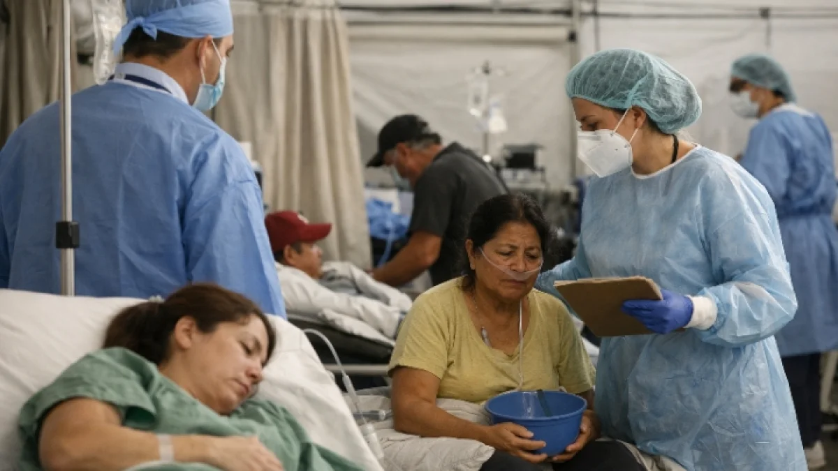
Seasonal influenza remains one of the world's most persistent infectious diseases. According to the World Health Organization, seasonal outbreaks cause millions of cases of severe illness and hundreds of thousands of deaths globally each year. Although vaccines and antiviral drugs have reduced the burden, influenza viruses continually evolve, forcing scientists to update vaccines and search for new therapeutic approaches.
For Jan Kosinski, a group leader at EMBL Hamburg and one of the study's senior authors, the challenge has long been that researchers could identify many viral and human proteins but struggled to determine how they actually interacted during infection.
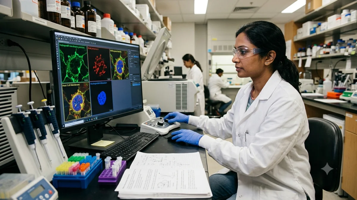
The team's solution combined in-cell cross-linking mass spectrometry with computational structural modeling, including a modified version of AlphaFold, allowing researchers to observe protein contacts where they naturally occur inside infected cells. Rather than reconstructing interactions after cells had been broken apart, the method captured molecular relationships while infection was actively unfolding.
The resulting map revealed two findings that particularly caught the researchers' attention.
One involved hemagglutinin, the protein that covers the virus's surface and enables influenza to enter human cells. Scientists found previously unrecognized human proteins involved in folding and processing hemagglutinin as it moved through the cell's protein-manufacturing system, offering possible new targets for future antiviral research.
The second finding centered on tiny structures inside the cell nucleus known as paraspeckles.
These membrane-free compartments normally help organize RNA and contribute to cellular stress responses. During influenza infection, however, the researchers observed paraspeckles consistently breaking apart across multiple human cell lines and different influenza strains. Their disruption released RNA-binding proteins that the virus appears to exploit to support its own replication.
"What surprised us most was the paraspeckles," first author Iuliia Kotova, now at ETH Zurich after completing doctoral work at EMBL Hamburg, said in a statement released by EMBL. "Watching these tiny organelles in the nucleus dissolve, consistently across every cell line and every flu strain we tested, told us this isn't a side effect of infection—it might be a strategy."
Kosinski said the findings suggest the virus may gain two advantages simultaneously: accessing proteins that promote viral replication while potentially weakening parts of the cell's antiviral defense system.
For researchers working on antiviral medicines, such molecular detail matters because many existing influenza drugs target viral proteins that can mutate over time. Human proteins or cellular processes essential for viral replication may prove more stable therapeutic targets, although translating laboratory discoveries into approved medicines typically requires years of additional research.
The study also illustrates how advances in structural biology have transformed virology since the COVID-19 pandemic accelerated investment in technologies capable of visualizing biological processes at increasingly fine resolution.
Boris Bogdanow, now a junior research group leader at Charité–Universitätsmedizin Berlin and a co-author of the study, said the experimental workflow was designed to capture fleeting protein interactions that earlier methods often missed.
According to Bogdanow, the approach could eventually be applied beyond seasonal influenza to viruses with greater pandemic potential, including highly pathogenic avian influenza strains such as H5N1.
That broader application reflects a continuing lesson from infectious disease research: understanding familiar viruses often prepares scientists for future threats. The techniques developed while studying one pathogen frequently become tools for investigating another.
The research also depended on collaboration across multiple institutions, with scientists contributing expertise in proteomics, structural biology, microscopy and computational modeling. Such multidisciplinary efforts have become increasingly common as modern biology tackles questions too complex for any single laboratory to answer alone.
Outside research laboratories, the immediate reality of influenza remains unchanged. Vaccination continues to be the primary method of preventing severe disease, while antiviral medicines remain most effective when administered early in infection.
Yet every improvement in understanding the virus's biology adds another piece to a puzzle scientists have been assembling for decades.
Rather than depicting influenza as an invisible enemy, the new molecular map portrays it as an opportunistic intruder—one that succeeds by redirecting the machinery already present inside human cells. By revealing those interactions with unprecedented clarity, researchers hope future generations of antiviral medicines may interrupt the process before the virus gains control.